

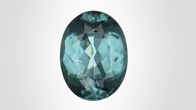
The Tokyo laboratory recently received a 0.70 ct blue-green oval modified brilliant measuring 6.62 × 4.99 × 3.10 mm (figure 1). Standard gemological testing yielded a refractive index of 1.620–1.640, uniaxial interference pattern, and a hydrostatic specific gravity of 3.10, all consistent with tourmaline. No Cu was detected with energy-dispersive X-ray fluorescence (EDXRF). UV-Vis-NIR spectrometry did not show Cu absorption bands but did show a strong Fe absorption. This stone, therefore, did not meet the requirement to be designated as a Paraíba tourmaline, whose dominant coloring agent of green/blue must be Cu (LMHC Information Sheet #6, 2012).
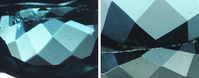
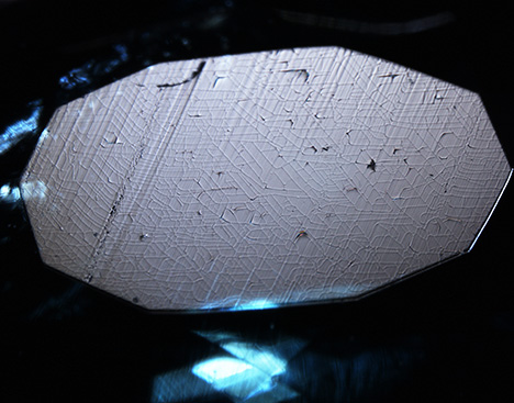
Microscopic observation revealed a unique networked pattern, resembling the pattern of a circuit board, covering the overall surface of the stone. The surface features mainly pointed in three directions on the crown but did not show a clear three-directional pattern on the pavilion (figure 2). Viewed under reflected light, some features at the table facet were parallel to the “polish lines” (figure 3). No evidence of coating and no color concentration along the pattern were found. The conoscope image with crossed polarized filters showed that the table facet was perpendicular to the c-axis. Because tourmaline belongs to the trigonal crystal system, the features appeared to be crystallograpically aligned. That could also explain why the patterns were different between the crown and the pavilion.
We also observed tension cracks surrounding fluid inclusions, which indicated the possibility of heat treatment. In addition, this color of tourmaline is commonly heat treated to improve its color.
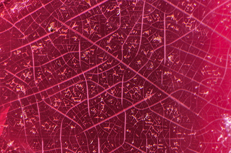
Because the unique patterns were related to the crystal structure, they may have been caused by thermal shock related to heat treatment and/or acid etching. Similar crystallographically aligned surface features have been found on rough crystals of Tanzanian tourmaline and Burmese spinel (figure 4; N. Renfro, pers. comm., 2021). What caused this pattern and how it came to be revealed on a faceted stone remains a mystery. More samples and further experiments are needed to unravel the cause of these unique and interesting patterns.
The Tokyo laboratory recently received a 0.70 ct blue-green oval modified brilliant measuring 6.62 × 4.99 × 3.10 mm (figure 1). Standard gemological testing yielded a refractive index of 1.620–1.640, uniaxial interference pattern, and a hydrostatic specific gravity of 3.10, all consistent with tourmaline. No Cu was detected with energy-dispersive X-ray fluorescence (EDXRF). UV-Vis-NIR spectrometry did not show Cu absorption bands but did show a strong Fe absorption. This stone, therefore, did not meet the requirement to be designated as a Paraíba tourmaline, whose dominant coloring agent of green/blue must be Cu (LMHC Information Sheet #6, 2012).
Microscopic observation revealed a unique networked pattern, resembling the pattern of a circuit board, covering the overall surface of the stone. The surface features mainly pointed in three directions on the crown but did not show a clear three-directional pattern on the pavilion (figure 2). Viewed under reflected light, some features at the table facet were parallel to the “polish lines” (figure 3). No evidence of coating and no color concentration along the pattern were found. The conoscope image with crossed polarized filters showed that the table facet was perpendicular to the c-axis. Because tourmaline belongs to the trigonal crystal system, the features appeared to be crystallograpically aligned. That could also explain why the patterns were different between the crown and the pavilion.
We also observed tension cracks surrounding fluid inclusions, which indicated the possibility of heat treatment. In addition, this color of tourmaline is commonly heat treated to improve its color.
Because the unique patterns were related to the crystal structure, they may have been caused by thermal shock related to heat treatment and/or acid etching. Similar crystallographically aligned surface features have been found on rough crystals of Tanzanian tourmaline and Burmese spinel (figure 4; N. Renfro, pers. comm., 2021). What caused this pattern and how it came to be revealed on a faceted stone remains a mystery. More samples and further experiments are needed to unravel the cause of these unique and interesting patterns.


