

The author recently examined a batch of oil-bearing quartz crystals from Madagascar. Microscopic observation revealed morphologically diverse multicolored fluorescent inclusions. Raman spectroscopy, infrared spectroscopy, and X-ray diffraction analyses confirmed that the fluorescence originated from hydrocarbon compounds.
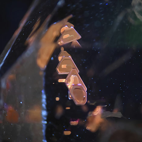
A set of inclined inclusions exhibited orange-red fluorescence under long-wave (365 nm) UV light (figure 1). Further examination revealed their distinct polygonal forms arranged in a regular, parallel orientation. Unlike protogenetic inclusions, these inclusions exhibited a highly ordered structure typical of negative crystals. Combined with microscopic observations, these inclusions resembled crimson terraced fields within the crystal, which ranged from triangles to pentagons.
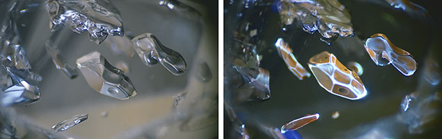
Another set of elongated negative crystals formed a pattern resembling a school of fish (figure 2). A compressed bubble observed in one of the negative crystals, which was inert under long-wave UV light, was reminiscent of a fish eye. While the negative crystals exhibited orange-red fluorescence, their crystal edges displayed bluish white fluorescence, suggestive of the shimmering luster of fish scales. This combination of vibrant fluorescent colors reveals the internal complexity of inclusions that otherwise appear homogeneous and monotone.
Yecheng Lin is affiliated with Xiamen Xinchengxin Trading Co., Ltd. in China.
The author recently examined a batch of oil-bearing quartz crystals from Madagascar. Microscopic observation revealed morphologically diverse multicolored fluorescent inclusions. Raman spectroscopy, infrared spectroscopy, and X-ray diffraction analyses confirmed that the fluorescence originated from hydrocarbon compounds.
A set of inclined inclusions exhibited orange-red fluorescence under long-wave (365 nm) UV light (figure 1). Further examination revealed their distinct polygonal forms arranged in a regular, parallel orientation. Unlike protogenetic inclusions, these inclusions exhibited a highly ordered structure typical of negative crystals. Combined with microscopic observations, these inclusions resembled crimson terraced fields within the crystal, which ranged from triangles to pentagons.
Another set of elongated negative crystals formed a pattern resembling a school of fish (figure 2). A compressed bubble observed in one of the negative crystals, which was inert under long-wave UV light, was reminiscent of a fish eye. While the negative crystals exhibited orange-red fluorescence, their crystal edges displayed bluish white fluorescence, suggestive of the shimmering luster of fish scales. This combination of vibrant fluorescent colors reveals the internal complexity of inclusions that otherwise appear homogeneous and monotone.
Yecheng Lin is affiliated with Xiamen Xinchengxin Trading Co., Ltd. in China.


